Release date:
2026-03-19 View count: 15
OMIP-015 offers a highly efficient and comprehensive panel for the investigation of human regulatory T cells (Tregs) and activated T cells, without the need for intracellular staining. This breakthrough design has significant implications for the study of T cell biology, particularly when considering the challenges often associated with intracellular marker use, such as fixation and permeabilization.
1. OMIP-015 Panel
|
Target
|
Fluorochrome
|
Function
|
|
Dead Cells
|
AqBlu
|
Exclude dead cells
|
|
CD3
|
BV785
|
T cell lineage
|
|
CD4
|
QD605
|
|
CD8
|
QD585
|
|
CD25
|
PE-Cy5
|
Treg
|
|
CD127
|
APC-eF780
|
|
CD39
|
PE-Cy7
|
Treg functionality
|
|
CD73
|
PE
|
|
CD38
|
PE-CF594
|
Other activation/ differentiation
|
|
CD45RA
|
QD705
|
|
CD45RO
|
FITC
|
|
HLA-DR
|
Alexa 680
|
|
PD-1
|
BV421
|
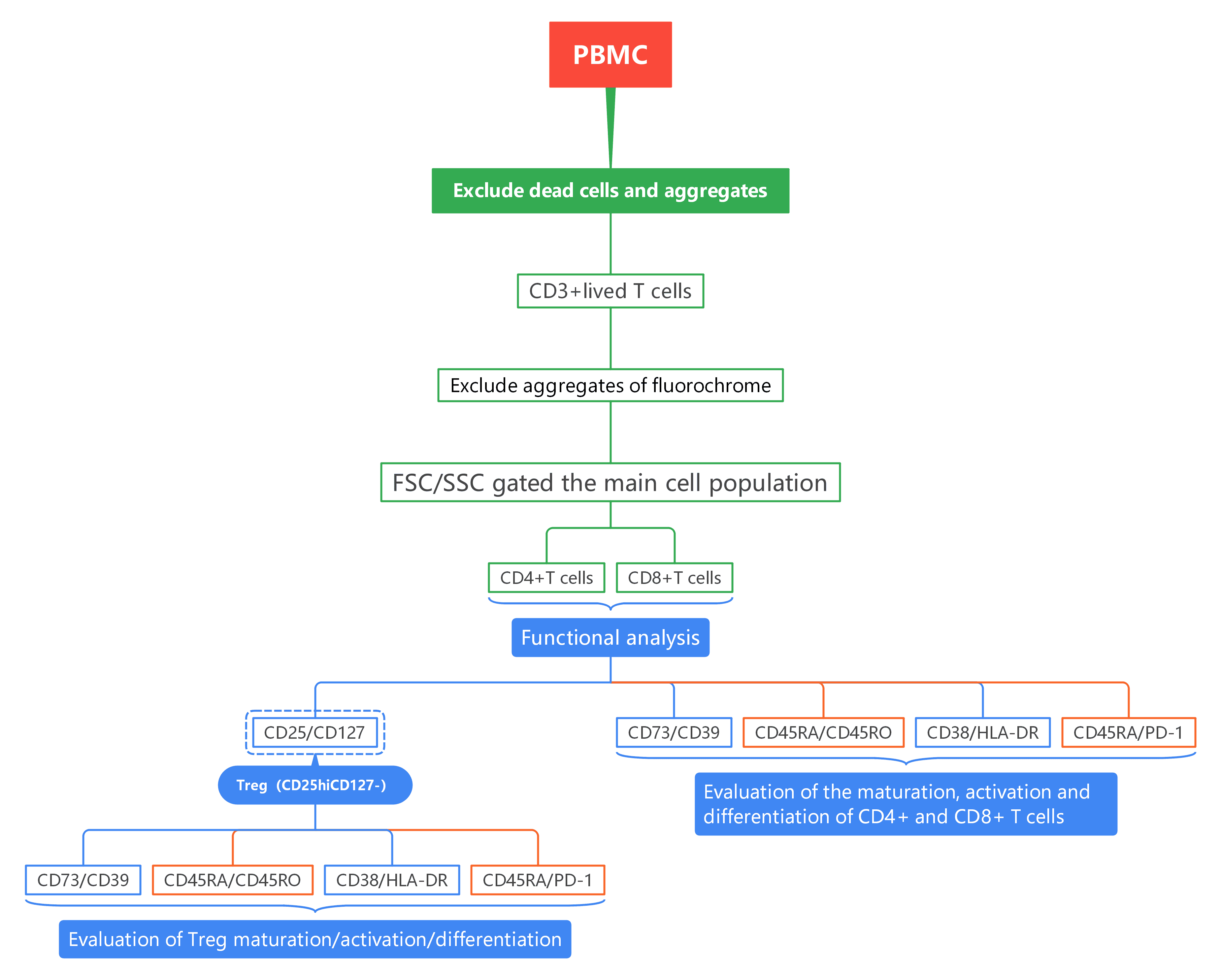
2. Gate Logic
3. Experimental Results
1). Excludeaggregates, and gated live CD3+ cells. Further, non-specific signals caused by dye aggregates are excluded using combinations of markers, and CD4+ and CD8+ T cells are subsequently isolated.
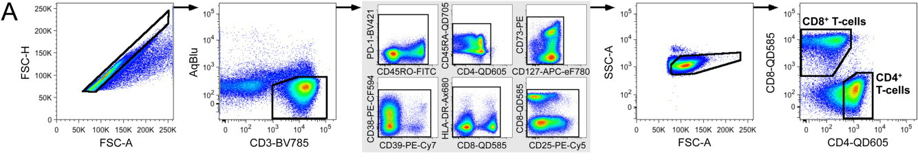
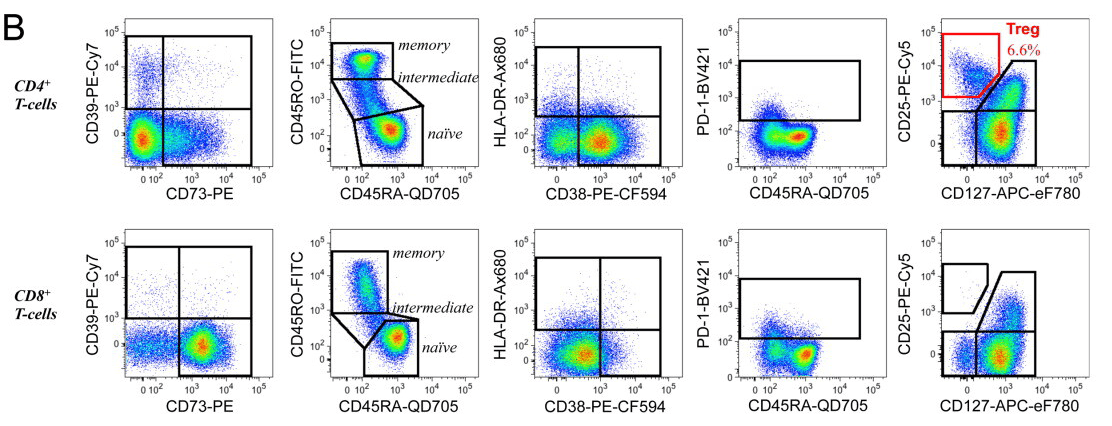
2). Within CD4+ and CD8+ T cells, analyzed the differentiation/activation abilities of cells using CD39/CD73, CD45RA/CD45RO, CD38/HLA-DR and CD45RA/PD-1, CD25/CD127 was used to determine Treg cells (CD25hiCD127-).
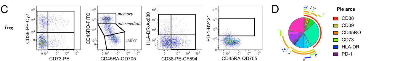
3). Further analysis of Treg cells investigated their maturation status, regulatory potential, and cell activation status using CD39/CD73, CD45RA/CD45RO, CD38/HLA-DR, and CD45RA/PD-1. Figure D shows the proportion of total Treg cells expressing any combination of the measured activation markers.
4. Protocol Interpretation
1). Simplified Experimental Workflow:
One of the most notable strengths of OMIP-015 is its ability to characterize Tregs and activated T cells without requiring intracellular staining. Traditionally, FoxP3, a canonical marker for Tregs, has been used for intracellular staining, which complicates the process due to fixation, permeabilization, and the potential for introducing experimental artifacts. OMIP-015 avoids this step entirely, simplifying the workflow and reducing the time and complexity of experiments. This approach also ensures that the panel is potentially compatible with live cell sorting, which is a significant advantage for subsequent functional or genomic analysis.
2). Innovative Use of Surface Markers:
OMIP-015 employs an innovative combination of surface markers to define Tregs and activated T cells. Tregs are identified through a combination of CD25 (IL-2Ra) and CD127 (IL-7Rα), with the Tregs being CD25^hi CD127^−. This avoids reliance on intracellular FoxP3, which has limitations in detecting subsets of Tregs in certain experimental conditions. The addition of functional markers like CD39 and CD73 enhances the panel's ability to differentiate between various Treg subsets with immunosuppressive functions. These markers are particularly useful for understanding the functional capacity of Tregs in regulating immune responses.
Moreover, CD39 and CD73, together with CD45RA and CD45RO, offer insights into the differentiation and activation status of Tregs. This is a critical feature for characterizing Tregs in various immune conditions and diseases, providing valuable information beyond mere phenotype identification.
3). Comprehensive Activation Marker Profile:
The inclusion of CD38, HLA-DR, and PD-1 adds an additional layer of depth to the analysis of T cell activation. These markers have been shown to correlate with disease control, particularly in HIV-1 infection, where CD38 and HLA-DR expression on T cells can provide insights into the activation and exhaustion states of these cells. By using these activation markers, OMIP-015 can help researchers gain a better understanding of the activation dynamics of Tregs and other T cell subsets under different immune conditions.
4). Relevance for Disease Research:
OMIP-015's ability to detect subtle differences in Treg subsets, particularly in the context of immune suppression and activation, holds great promise for research into autoimmune diseases, cancer immunotherapy, and infectious diseases such as HIV. Its potential for live cell sorting further enhances its applicability to functional studies, where the behavior of specific T cell subsets can be analyzed in greater detail.
5. Conclusion
This panel provides a streamlined and highly effective method for studying Tregs and activated T cells, enabling detailed characterization without the complexity of intracellular staining. The combination of surface markers for phenotype, activation, and function offers researchers a powerful tool for investigating T cell biology, with broad applications in both basic and translational immunology.
References:
[1] Mahnke YD, Beddall MH, Roederer M. OMIP-015: human regulatory and activated T-cells without intracellular staining. Cytometry A. 2013 Feb;83(2):179-81.
About Us
As a strategic venture of AtaGenix (established 2011), abinScience was founded in 2023 to deliver premium life science reagents that accelerate discovery. Our flow cytometry antibody products cover commonly used detection markers, with a wide variety to meet the research needs of multiple species (Human, Mouse, Rat, Dog, Hamster, Monkey, etc.). We provide stable and reliable support for scientific research. For more information on abinScience flow cytometry antibodies, please click:
abinScience Flow Cytometry (FACS) Antibodies

 中文
中文 English
 한국어
한국어 日本語
日本語 Español
 Français
 Русский


