A 15-Colour Panel for Comprehensive Profiling of Human Regulatory T Cells
OMIP-004 was tailored to address the need for in-depth, multi-faceted analysis of human regulatory T cells (Tregs) across healthy and disease states. This optimized 15-colour flow cytometry panel integrates core Treg identifiers, functional markers, subset differentiation labels, and essential gating/activation tools, enabling simultaneous quantitative enumeration and detailed phenotypic characterization of Tregs in a single assay—with reliable performance on fresh, cryopreserved PBMCs, and ACK-lysed peripheral blood.
Technical Challenges in Treg Biology and Translational Research
Unraveling the phenotypic heterogeneity, functional diversity, and pathological relevance of Tregs is critical for advancing understanding of immune homeostasis, autoimmune disease mechanisms, and the efficacy of immunotherapies. However, conventional Treg analysis approaches often rely on a narrow set of canonical markers (e.g., FoxP3, CD25, CD127) and fail to capture the full spectrum of Treg subsets (naive/memory) or their suppressive capacity. Such fragmented profiling requires multiple experimental runs with separate panels, increasing procedural burden, compromising data consistency, and hindering holistic investigations of Treg-mediated immune regulation in both basic and clinical settings.
Core Design and Innovation of OMIP-004
OMIP-004’s true innovation lies in its systematic integration of Treg research demands, rather than basic experimental operations. Its 15-colour panel breaks the “single-dimension” limitation of traditional panels (only identifying FoxP3+CD25+ cells) by building a “phenotype-subset-function” three-dimensional marker system: it combines core Treg identifiers (CD25high, FoxP3, CD127low) with subset markers (CD45RA/CD197 to distinguish naive/memory Tregs) and functional indicators (CD39 to reflect suppressive activity), enabling one-step clarification of Treg heterogeneity. More importantly, it establishes a standardized staining and detection protocol applicable to fresh PBMCs, cryopreserved samples, and ACK-lysed peripheral blood—solving the problem that traditional panels often require protocol adjustments for different sample types (leading to poor data comparability).
Regulatory T cells (Tregs) are pivotal in maintaining immune homeostasis and modulating disease progression. However, their low frequency and phenotypic complexity present significant challenges for precise analysis. OMIP-004 addresses this by enabling multidimensional characterisation of Tregs through the integrated assessment of FoxP3, CD25, CD127, and CD39, alongside markers associated with differentiation and function. This allows researchers to simultaneously determine Treg subset distribution and functional attributes, providing a robust tool for gaining deeper insights into the role of Tregs in health and disease.
1. OMIP-004 Panel
2. Experimental Results and Gate Logic
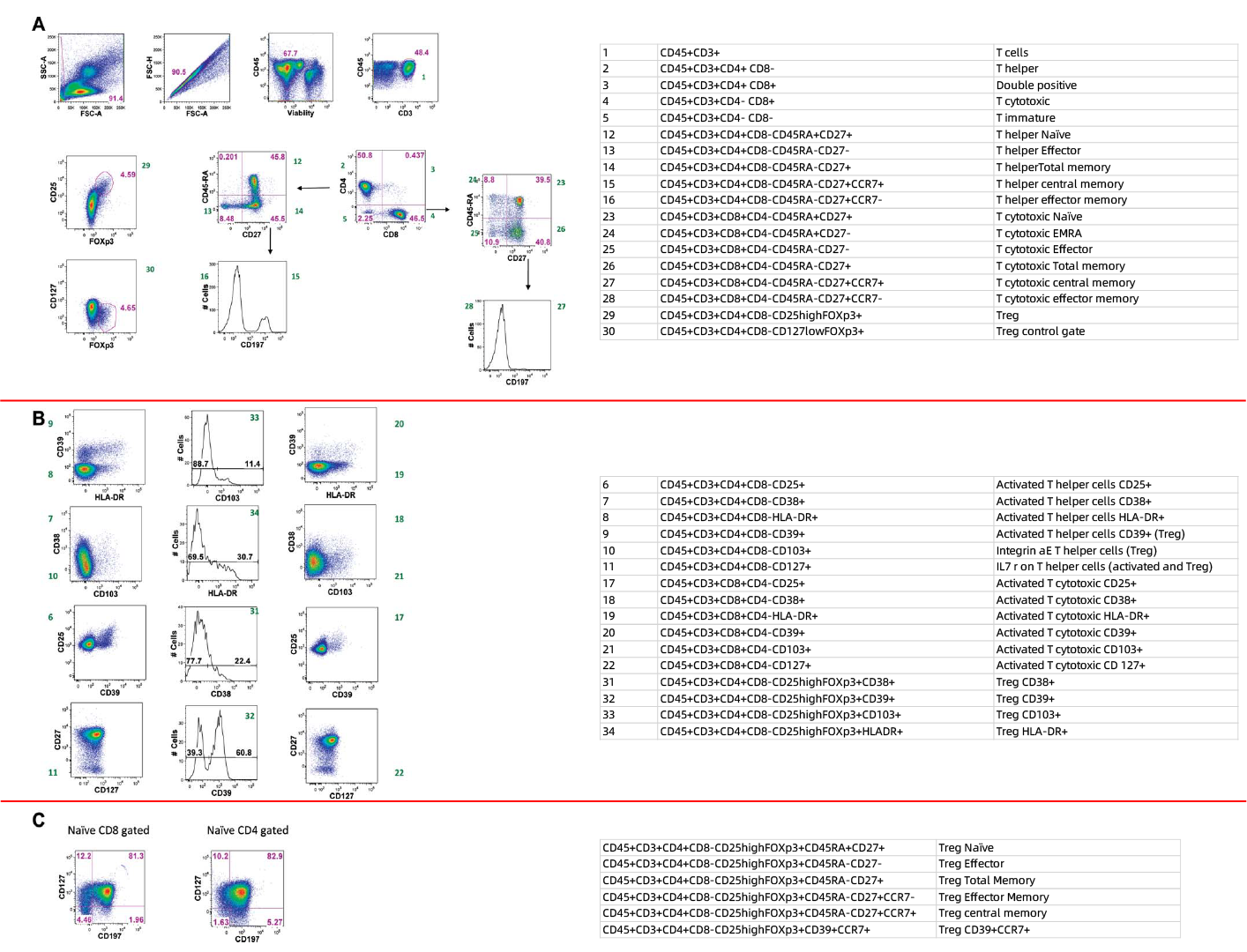
A
Stepwise Screening and Identification of Treg Cell Candidate Populations
Exclude cell aggregates, dead cells. Then, T cells were divided into CD4+ helper T cell and CD8+ cytotoxic T cell subsets using CD3/CD4/CD8 markers. Among CD4+ T cells, Treg cells were gated using CD25/CD127/FoxP3. Meanwhile, the differentiation status of CD4+ helper T cells and CD8+ cytotoxic T cells was analyzed via CD45RA/CD27/CD197.
B
Differences in Marker Expression Between Tregs and Other T Cell Subsets
Compare the marker expression of CD8+ T cells (left), Treg candidates (middle) and conventional CD4+ T cells (right): Tregs exhibited high CD39 (linked to strong suppressive activity)/CD25 and low CD127, which matched the reported core phenotype (CD25high, FoxP3+, CD127low) and confirmed their functional specificity. Expressions of CD38, HLA-DR (activation markers) and CD103 (homing marker) aided in evaluating Treg activation and localization potential, with green values defining phenotypic boundaries.
C
Molecular Characteristics of Naive T Cells and Stage Classification of Treg Subsets
Analyze the expression of CD197 (CCR7) and CD127 in naive CD8+ (left) and CD4+ T cells (right), with naive T cells defined by CD45RA+CD27+ (per Figure 1A gating): Tregs within naive CD4+ T cells retained CD127low/CD197high features (suggesting lymph node homing potential). Marker expression differences between the two naive subsets provided a molecular basis for distinction, with green values corresponding to right-side table data for subset proportion analysis.
3. Panel Interpretation
3.1 Comprehensive Treg Characterisation
Unlike earlier T-cell panels, OMIP-004 is specifically designed for in-depth immunophenotyping of human Tregs. It moves beyond conventional reliance on CD25high and FoxP3 by incorporating CD127low and the functionally relevant marker CD39, thereby achieving highly specific and high-resolution identification of Tregs.
3.2 Integrated Analysis of Differentiation and Functional States
The panel concurrently includes differentiation markers (CD45RA, CD27, CCR7) and functional/activation markers (CD39, HLA-DR, CD38, CD103). This enables the simultaneous discrimination of naïve versus memory Treg subsets and evaluation of their activation status and suppressive potential within a single sample, yielding rich data for functional studies.
3.3 Complementary and Progressive to OMIP-001
Building upon the T-cell memory subset framework established in OMIP-001, OMIP-004 incorporates additional Treg-specific markers. This facilitates a layered analysis, allowing researchers to progress seamlessly from overall T-cell immunophenotyping to memory differentiation, and finally to detailed Treg phenotype and function, all within a consistent analytical structure.
4. Applications
In-depth Treg characterization Autoimmune disease research Immunotherapy monitoring Treg subset analysis Immunosuppression assessment
OMIP-004 is a critical tool in immunological and clinical research, enabling detailed exploration of human Treg phenotypes and subset distributions, evaluation of their immunosuppressive potential, and supporting investigations into autoimmune diseases as well as the monitoring of immunotherapy efficacy, with applicability in both healthy and disease contexts.
5. Conclusion
OMIP-004 establishes a high-dimensional flow cytometry panel for the detailed dissection of human Tregs. It accurately identifies Tregs using core markers (CD25high, FoxP3, CD127low) and enables refined subpopulation analysis through the inclusion of functionally relevant molecules (CD39, HLA-DR, CD38, CD103) and differentiation markers (CD45RA, CD27, CCR7). The panel is suitable for use with fresh or cryopreserved PBMCs and whole blood, reliably detecting rare Treg populations and providing information on their differentiation status and suppressive function. OMIP-004 serves as a high-resolution tool for investigating Treg phenotype and function. It complements broader T-cell panels such as OMIP-001, supporting a continuous analytical workflow from general T-cell profiling to specific Treg subset characterisation. This panel holds considerable value for research in autoimmunity, cancer immunology, and immune regulation.
Get OMIP-004 Compatible Flow Cytometry Antibodies
abinScience provides validated flow cytometry antibodies covering key targets in this panel, supporting your research on Treg cells
References
[1] Biancotto A, Dagur PK, Fuchs JC, Langweiler M, McCoy JP Jr. OMIP-004: in-depth characterization of human T regulatory cells. Cytometry A. 2012 Jan;81(1):15-6.

About Us
As a strategic venture of AtaGenix (established 2011), abinScience was founded in 2023 to deliver premium life science reagents that accelerate discovery. Our flow cytometry antibody products cover commonly used detection markers, with a wide variety to meet the research needs of multiple species (Human/Mouse/Rat/Dog/Hamster/Monkey, etc.). We provide stable and reliable support for scientific research.
Explore abinScience Flow Cytometry Antibodies

 中文
中文 English
 한국어
한국어 日本語
日本語 Español
 Français
 Русский