Release date:
2025-12-05 View count: 255
In traditional flow cytometry-based B cell subset identification, researchers typically rely on a limited number of markers such as IgD, CD27, CD38, and CD24 to classify B cell subsets. However, this approach has significant limitations: on the one hand, key markers are limited, making it difficult to distinguish major subsets clearly; on the other hand, it fails to integrate multiple co-expression markers within a single panel, potentially overlooking novel subsets. Additionally, repeated staining increases sample consumption and is less efficient for studying rare samples.
To address these challenges, OMIP-003 has developed a 12-colour panel that integrates anchor markers (CD19, CD3, Aqua viability dye, IgD, CD24, CD27, CD38) with extended markers (CD21, CD95, CD45/B220, CXCR3, 9G4 antibody). This panel not only standardizes the classification of core B cell subsets but also reveals the activation state, autoreactivity, and homing potential of memory B cells.
1. OMIP-003 Panel
|
Target
|
Fluorochrome
|
Function
|
|
CD3
|
Pacific Blue
|
Excluding T cells
|
|
CD19
|
APC-Cy7
|
The main markers of B cells, used for initial screening of live B cells
|
|
Dead cells
|
Aqua
|
Live/dead dyes
|
|
IgD
|
FITC
|
Distinguishing between naive B cells (IgD+) and memory B cells (IgD-)
|
|
CXCR3
|
PE
|
Chemokinetic receptors, reflecting the ability of B cells to migrate to inflamed tissues
|
|
CD24
|
PE-Alexa 610
|
High expression suggests naive or immature B cells, low expression suggests mature/activated B cells
|
|
CD21
|
PE-Cy5
|
CR2, low expression (CD21^low) suggests activated or potentially autoimmune-associated B cells
|
|
CD38
|
PerCP-Cy5.5
|
Labeling plasmablasts (CD27+CD38+) in the IgD- region
|
|
CD45/B220
|
PE-Cy7
|
Aiding in distinguishing between mature and immature/germinal center B cells
|
|
CD27
|
Qdot605
|
Distinguishing between converted memory (CD27+) and CD27- memory B cells
|
|
CD95
|
APC
|
Fas receptor, upregulation indicates B cell activation or apoptosis-proneness
|
|
VH4-34 idiotype
|
Biotin- Alexa 680
|
VH4-34-encoded autoantibody B cell markers, mainly found in the IgD+ naïve region in healthy individuals
|
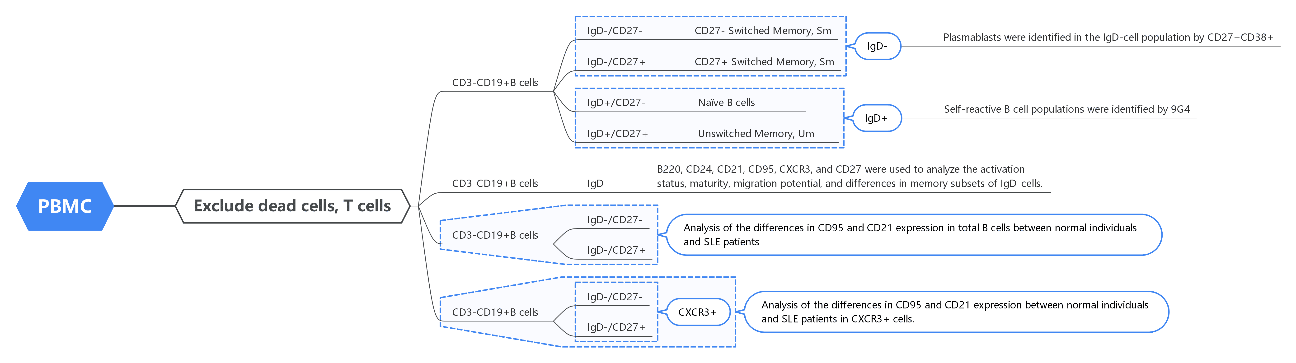
2. Gate Logic
3. Experimental Results
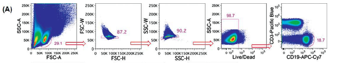
1). PBMC Analysis:
Gated the main cell populations, exclude aggregates, and gated live cells. B cells are identified by CD3-CD19+ .
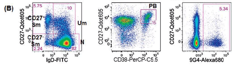
2). B Cell Subsets:
Within the B cell population, IgD/CD27 differentiation is used to classify B cells at different stages of differentiation. Plasmablasts are identified as CD27+CD38+ in the IgD- population. Autoreactive B cells are marked by 9G4 in the IgD+ population.
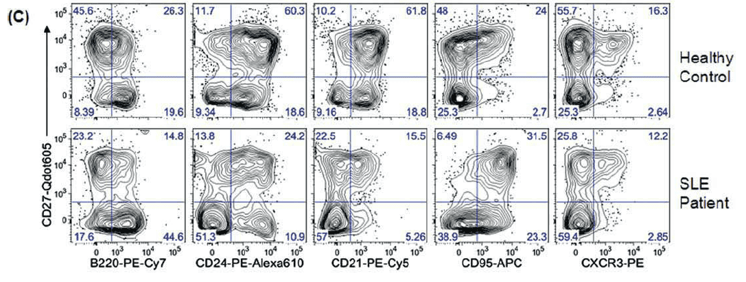
3). Activation, Maturity, and Migration:
In the IgD- subset, the expression of B220, CD24, CD21, CD95, CXCR3, and CD27 is used to assess activation status, maturity, migration potential, and memory subgroup differences, comparing healthy individuals with SLE patients.
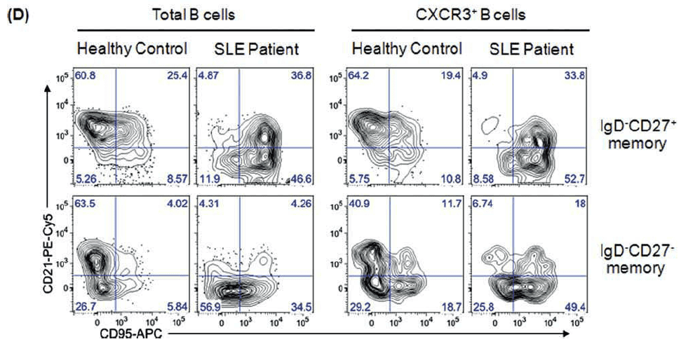
4). Memory B Cells in SLE:
Comparison of the CD3-CD19+ B cell population and CD3-CD19+CXCR3+ subsets reveals differences in CD95/CD21 expression between IgD-CD27- and IgD-CD27+ memory cells.
4. Protocol Interpretation
1). Comprehensive Coverage of Human Memory B Cell Subsets
OMIP-003 panel enables the differentiation of naïve B cells (IgD+CD27-), unconverted memory B cells (IgD+CD27+), and converted memory B cells (CD27+ SM and CD27- SM) in a single experiment. Additionally, plasmablasts (CD27+CD38+) are also identified. This design overcomes the limitations of traditional approaches, providing a standardized analysis of major B cell subsets within the same panel.
2). Integration of Function and Migration States
CD21 and CD95: Used to determine B cell activation. In SLE patients, CD27- SM cells frequently exhibit downregulation of CD21 and upregulation of CD95, indicating an aberrant activation state.
CXCR3: Used to assess B cell migration potential to inflammatory tissues. Upregulation of CXCR3 in disease settings suggests that these B cells may be more likely to enter the inflammatory microenvironment.
CD24 and B220/CD45: Assist in distinguishing developmental stages and maturation status of B cells.
3). B Cell Differences Between SLE and Healthy States
In healthy individuals, the distribution of B cell subsets is relatively stable, with autoreactive B cells (9G4+) predominantly confined to the naïve region and not entering the memory compartment. In contrast, in SLE patients, there is a marked expansion of CD27- SM B cells, accompanied by abnormal activation and migration-related phenotypic changes, suggesting that this population may play a pivotal role in disease pathogenesis.
5. Conclusion
OMIP-003 establishes a standardized framework for phenotypic analysis of human memory B cells. It not only provides clear differentiation between traditional and novel memory B cell subsets but also integrates dimensions of activation, migration, and autoreactivity for comprehensive interpretation. This panel is a milestone tool in memory B cell research, with significant applications in autoimmune studies, immunology of infection, vaccinology, and transplantation immunology.
References:
[1] Wei C, Jung J, Sanz I. OMIP-003: phenotypic analysis of human memory B cells. Cytometry A. 2011 Nov;79(11):894-6. doi: 10.1002/cyto.a.21112. Epub 2011 Jul 27.

About Us
As a strategic venture of AtaGenix (established 2011), abinScience was founded in 2023 to deliver premium life science reagents that accelerate discovery. Our flow cytometry antibody products cover commonly used detection markers, with a wide variety to meet the research needs of multiple species (Human, Mouse, Rat, Dog, Hamster, Monkey, etc.). We provide stable and reliable support for scientific research. For more information on abinScience flow cytometry antibodies, please click:

 中文
中文 English
 한국어
한국어 日本語
日本語 Español
 Français
 Русский


